
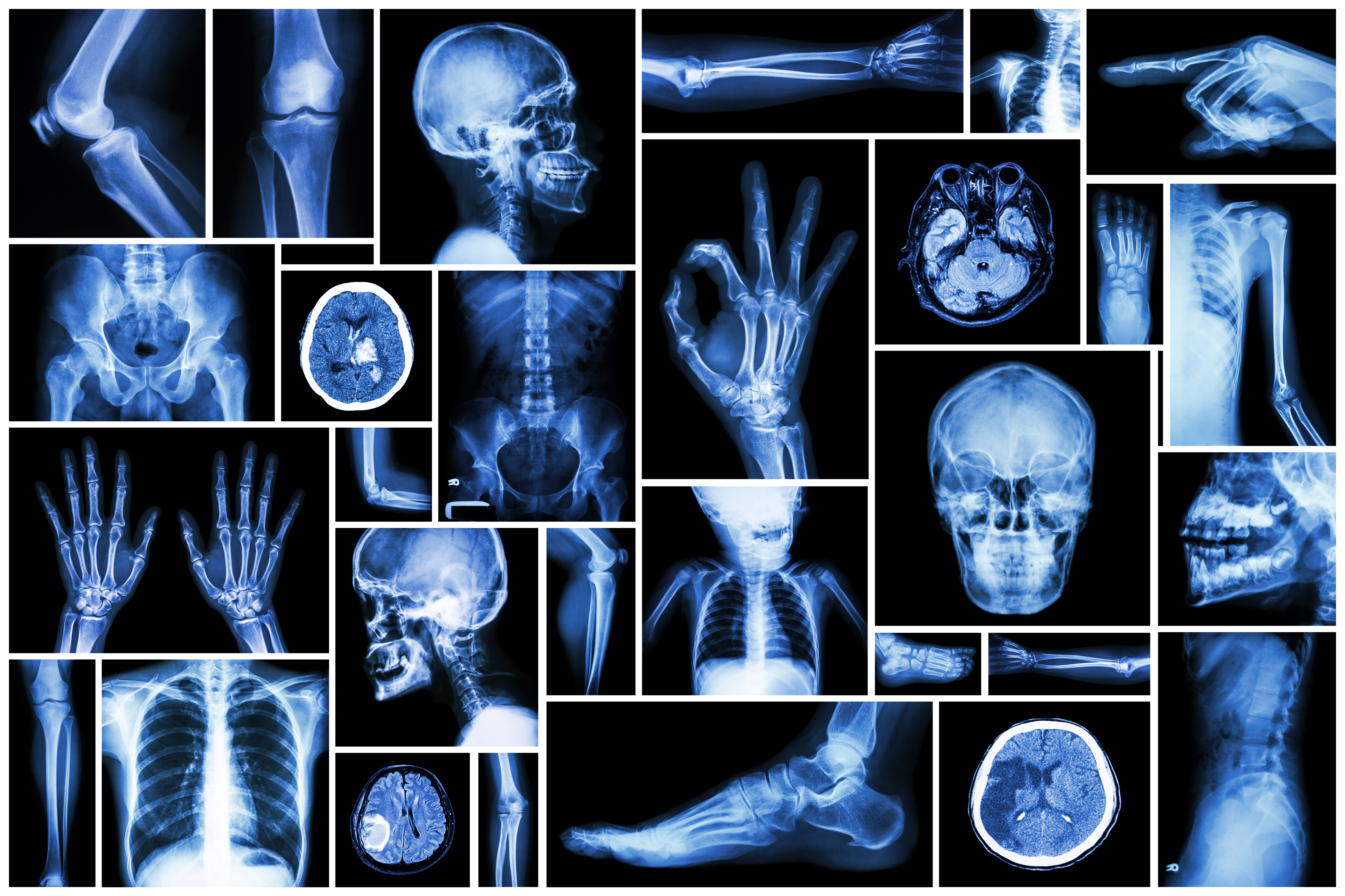
X-ray imaging, a mainstay in radiology for nearly a century, remains a simple, quick and effective tool to assess various body systems, including major organs and bones. It is often used to identify broken bones, however, it can also assist with the diagnosis of problems in many other parts of the body. These include major organs such as the heart, lungs and intestines, to smaller structures within the ear, nose or throat.
There are many specific types of X-ray imaging that each carry their own name that are related to the area of the body or type of structure that is imaged. Some examples are listed below:
There are many specific types of X-ray imaging that each carry their own name that are related to the area of the body or type of structure that is imaged. Some examples are listed below:
| Exam Type Angiography Arthrography Chest X-ray Cholangiography Lymphangiography Mammography Myelography Myelography Skeletal X-ray Urography |
Body part/area Blood vessels Joints Chest, heart, lungs Bile ducts Gallbladder Lymph system Breasts Spinal canal Bones Kidneys, ureters and bladder |
In many cases, X-rays are enhanced by the use of contrast. Contrast is a substance that makes the area of interest show up better on the X-ray than surrounding tissues and structures, thereby making it easier to diagnose. The method of introducing contrast into the patient depends upon the area being studied, so it might be taken orally (swallowed), infused through a catheter or placed through an intravenous injection.
X-Rays are produced by bombarding a tungsten electrode inside an X-ray tube with a stream of electrons. The x-ray equipment will focus this energy into a beam which passes through the body and is collected on a piece of film or electronic detectors. Because the different tissues within the body are of different densities, those waves are attenuated (weakened) at differing rates as they pass through. Bone, for example, is very dense and absorbs a lot of the x-rays, while the tissues surrounding the bone are less dense and absorb less of the x-ray. It is this difference in the absorption of the waves that creates variations in the exposures are translated into an image that can be subsequently examined (interpreted) by a radiologist.
There are two major categories of x-ray imaging; radiographic that produces still images; and fluoroscopicthat produces moving pictures similar to an x-ray video. This latter type of x-ray is viewed on a television monitor.
Digital radiographic x-ray machines use techniques similar to traditional analog (or film) with the exception that the images are digitally acquired and sent to a computer for viewing. This technique produces images that can be easily reproduced to allow the physician to enhance and manipulate them, as well as store them electronically and share with other physicians. Digital x-ray also may use a lower dosage of radiation to create the same (or better) quality images as film.