
Ultrasound exams use high frequency sound waves to produce images of the soft tissues and organs within the body. One of the highly valuable characteristics of ultrasound is its inherent safety, as it does not make use of any ionizing radiation (such as x-rays do).
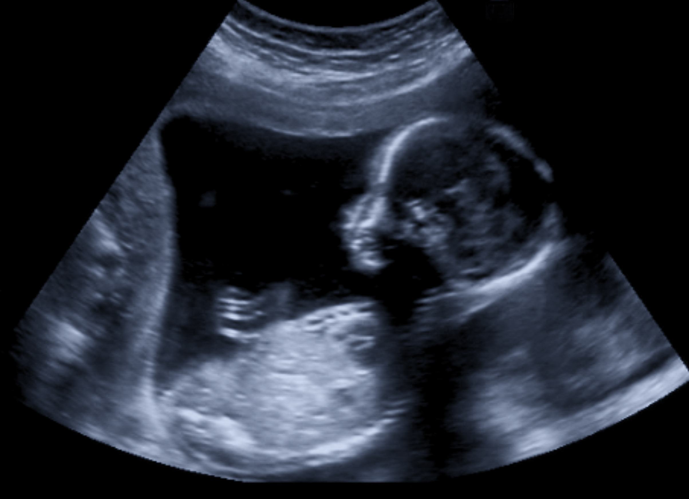
Ultrasound is used in obstetrics and gynecology to view the fetus or detect pelvic problems and abnormalities in the uterus or ovaries. Ultrasound can examine other soft tissue organs such as the heart, gallbladder, liver, pancreas, kidneys and bladder. It is often used to diagnose diseases of blood vessels in the neck and legs.
Ultrasound captures the echo of a sound wave as it is reflected back from a boundary and converts that data into an image. A hand-held probe that placed against your body transmits the sound waves. Although they are sound waves, you cannot hear them because they are beyond the frequency range of human hearing.
As the sound waves travel through the body they bounce off of tissue boundaries, such as bone or fluid, at differing rates. Some of the sound waves continue to pass on to the next tissue boundary. When the waves are reflected back, the probe detects the time delays and intensities of the echoes are processed into viewable images. A two-dimensional image is formed on the computer screen that can be viewed in real-time, meaning a moving picture is created which can display the motion of various structures. Blood flow and organ performance (such as the motion of the heart walls and valves) can be measured.