
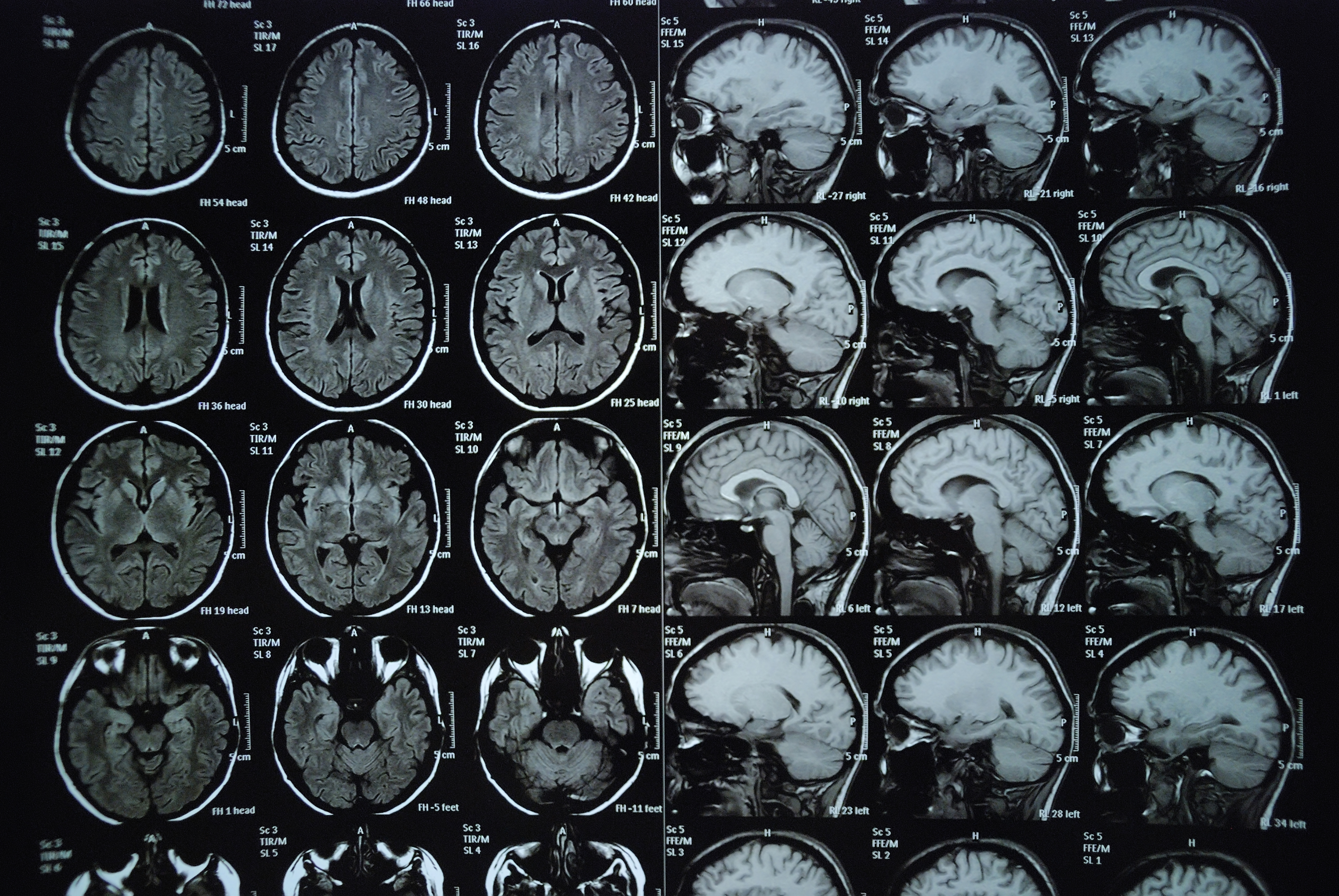
Magnetic Resonance Imaging (MRI) produces high-resolution images of the inside of the body differently than any other form of imaging. It produces two-dimensional and three-dimensional images that can identify very specific types of tissues. It is especially useful with accessing soft tissues such as nerves, as well as soft tissue that is in close proximity to boney structures, such as connective tissue in joints. In addition to showing tissue abnormalities, it can also be used to view blood flow within the body.
MRI is widely used for diagnosing or identifying tumors and masses, viewing torn ligaments or cartilage, diagnosing spine problems, strokes and multiple sclerosis (MS).
MRI technology uses a powerful magnet that affects the protons or hydrogen atoms within the body, forcing them to align to the magnetic field. Radiofrequency (RF) waves are then applied in pulses which cause hydrogen atoms. The atoms emit energy one the RF pulse stops which can be detected and processed into images. Because the amount of hydrogen varies for every substance, each type of tissue and fluid has a unique signature that can be transformed into very detailed images. This allows radiologists to more easily determine what is normal and what is not normal.
Regions of the body can be viewed from any angle without moving the patient, as is necessary with x-rays (and CT scans). Similar to ultrasound, MRI produces images without the use of ionizing radiation, making it very safe to use. Because of the strong magnetic fields employed, however, MRI’s cannot be performed on any patient with certain metal implants. Very careful screening and interview are conducted to understand a patient’s history before an MRI scan is performed.