
A fluoroscope produces a video (moving) x-ray. Continuous x-ray beams are used to view an organ or part of the body in real time. These images are viewed on a computer screen or television monitor. Fluoroscopy helps physicians to see catheters as they guide them into blood vessels, as with arteriography. More commonly, fluoroscopy is used to examine the stomach and bowels and their movements/function. It is also used to detect obstructions in airways and blood vessels. Contrast is often used during fluoroscopic studies.
Fluoroscopy is most often used to view the upper GI tract, which includes the stomach, esophagus, duodenum and the upper small intestine. It is also used to view the lower GI tract.
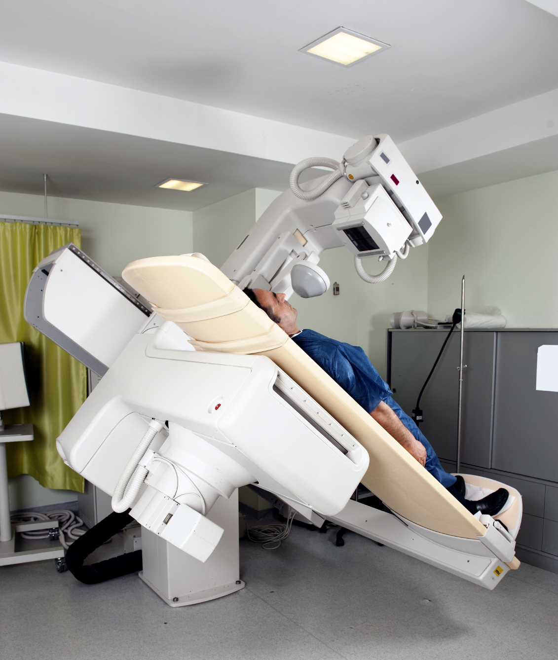
The fluoroscope is an x-ray machine that can use either a continuous or a pulsing x-ray beam that is captured by an image intensifier that increases the brightness of the image that it can be viewed on a display screen. This is connected to a video camera that produces a two-dimensional image on a video monitor. The camera output can be digitized by computers for enhancement and storage, as well.